TRYING TO CONCEIVE?
Pregnancy is one of the most awaited moment by a married couple in the world.
Sometimes there are some couples who can not quickly get it like other couples.
For such couples to be able to get pregnant sometimes, they have to labor harder and longer and often not within a reasonable time.

INFERTILITY
Infertility means you cannot get pregnant (conceive).
There are 2 types of infertility:
Primary infertility refers to couples who have not become pregnant after at least 1 year having sex without using birth control methods.
Secondary infertility refers to couples who have been able to get pregnant at least once, but now are unable.
Infertility: So what’s the difference between having trouble trying to get pregnant and being infertile?
Well, if the woman is under 34, she and her partner are considered infertile if they haven’t conceived after having 12 months of unprotected sex.
If she’s over 35, they’re considered infertile after six months of trying.
Causes of Infertility
Many physical and emotional factors can cause infertility. It may be due to problems in the woman, man, or both.
Female Infertility
Female infertility may occur when:
A fertilized egg or embryo does not survive once it attaches to the lining of the womb (uterus).
The fertilized egg does not attach to the lining of the uterus.
The eggs cannot move from the ovaries to the womb.
The ovaries have problems producing eggs.
Female infertility may be caused by:
~ Autoimmune disorders, such as antiphospholipid syndrome (APS)
~ Birth defects that affect the reproductive tract
~ Cancer or tumor
~ Clotting disorders
~ Diabetes
~ Drinking too much alcohol
~ Exercising too much
~ Eating disorders or poor nutrition
~ Growths (such as fibroids or polyps) in the uterus and cervix
~ Medicines such as chemotherapy drugs
~ Hormone imbalances
~ Obesity
~ Older age
~ Ovarian cysts and polycystic ovary syndrome(PCOS)
~ Pelvic infection or pelvic inflammatory disease(PID)
~ Scarring from sexually transmitted infection, abdominal surgery or endometriosis
~ SmokingSurgery to prevent pregnancy (tubal ligation) or failure of tubal ligation reversal (reanastomosis)
~ Thyroid disease
⏩ Male Infertility
Male infertility may be due to:
~ Decreased number of sperm
~Blockage that prevents the sperm from being released
~ Defects in the sperm
Male infertility can be caused by:
~ Birth defects
~ Cancer treatments, including chemotherapy and radiation
~ Exposure to high heat for prolonged periods
~ Heavy use of alcohol, marijuana, or cocaine
~ Hormone imbalance
~Impotence
~ Infection
~ Medicines such as cimetidine, spironolactone, and nitrofurantoin
~ Obesity
~ Older age
~ Retrograde ejaculation
~ Scarring from sexually transmitted infections, injury, or surgery
~ Smoking
~ Toxins in the environment
~ Vasectomy or failure of vasectomy reversal
IF YOU HAVE BEEN TRYING TO CONCEIVE FOR A YEAR AND YOU ARE UNABLE TO GET PREGNANT, PLEASE GO WITH YOUR SPOUSE TO DO SOME MEDICAL EXAMINATIONS OR FERTILITY TESTS. THE RESULTS OF THESE TESTS WILL SERVE AS A POINTER ON HOW TO GET STARTED ON WHAT YOU NEED TO DO
Remember: It takes two fertile people to conceive a baby
BELOW ARE SOME FERTILITY TESTS YOU CAN DO TO GET YOU STARTED ON YOUR JOURNEY TO PARENTHOOD
Preliminary Fertility Tests for TTC mums
The under listed reports usually gives an insight into a woman's reproductive health.
- Hormonal Assay
- Pelvic scan or USS
- High Vaginal Swab HVS
- Hysterosalpingogram HSG
✔ Hormonal assay gives insight into which hormone is high or low
~ FSH, LH, Estrogen & Prolactin testings are done on the third day of your menstrual period.
~ Progesterone testing is done on the 21st day of your menstrual cycle.
✔ Pelvic scan will show the obstruction in the pelvic, either Uterine fibroids, ovarian cysts, PCOS, Endometriosis, Pelvic inflammatory disease (chlamydia or gonorrhea)
✔ HVS ~ High Vaginal Swab (or Culture & sensitivity test) will show perhaps if it is Mild, scanty, moderate or heavy growth of a named isolated infection; eg Staphylococcus, E coli, Coliform, Chlamydia, Gonorrhea, Syphillis, Genital herpes, Bacterial Vaginosis BV, Cervicitis etc.
If there is infection in the report then I will examine: the Hysterosalpingogram ~ HSG
✔ HSG will show if the fallopian tube(s) is (are) patent; that is free or blocked. If there is a blockage, it will reveal which of the tubes, Left or Right that is blocked.


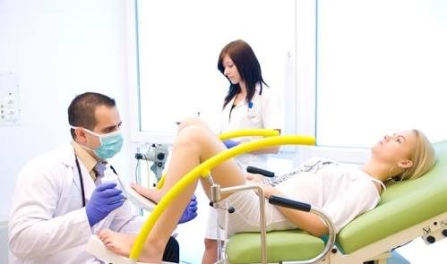
PELVIC OR GYNAECOLOGIC ULTRASOUND
Gynaecology is the branch of medicine that deals with women’s health issues, especially the study and management of female pelvic disorders.
A pelvic or gynaecologic ultrasound is an ultrasound of the female pelvis. It examines the female pelvic organs including the uterus (commonly called the “womb”), the endometrium (the lining of the uterus), the cervix and the ovaries.
HOW IS A PELVIC ULTRASOUND PERFORMED?
Most pelvic ultrasounds are performed using both the transabdominal and transvaginal approaches.
Transabdominal ultrasound involves scanning through your lower abdomen. Transabdominal ultrasound usually provides an overview of the pelvis rather than detailed images.
The transabdominal assessment is particularly helpful for the examination of large pelvic masses extending into the abdomen, which are not always well viewed with transvaginal ultrasound.
A small amount of ultrasound gel is put on the skin of the lower abdomen, with the ultrasound probe then scanning through this gel. The gel helps improve contact between the probe and your skin.

Transvaginal ultrasound
~ is an internal ultrasound. It involves scanning with the ultrasound probe lying in the vagina. Transvaginal ultrasound usually produces better and clearer images of the female pelvic organs, because the ultrasound probe lies closer to these structures.
The transvaginal ultrasound probe is thin, about 2cm diameter. The probe is covered with a disposable protective sheath. A small amount of ultrasound gel is placed on the end of this probe. The probe is then gently inserted a short distance into the vagina. All transvaginal probes have been cleaned and sterilised according to recommended protocols.
Your privacy will always be respected during a pelvic ultrasound, especially the transvaginal examination. You will have a towel covering your lower body, in addition to wearing a gown during the transvaginal ultrasound.
You will always have a choice about whether transvaginal ultrasound is performed. If you have concerns about transvaginal ultrasound, please discuss this with your sonographer before your pelvic ultrasound begins.

FEMALE HORMONE PROFILE OR ASSAY
The Female Hormone Profile is a hormone test for menstruating women or women that have not gone through the menopause.
The female hormones include FSH, LH, Prolactin, Estrogen and Progesterone, and levels of these hormones vary according to a certain pattern during the menstrual cycle.
If there is a problem with the hormonal cycle, then it can lead to problems such as irregular periods, heavy periods, absent periods, PMS, or infertility.
This test is valuable, giving us information about the hormonal cycle, which enables us to rationalise treatment in the best possible way.

High Vaginal Swab (HVS)
~ is a technique used in Obstetrics and Gynaecology to obtain a sample of discharge from the vagina.
This is then sent for culture and sensitivity.
It is commonly used to test for the presence of the following:
~ Candidiasis infection,
~ Bacterial Vaginosis BV
~ Trichomonas Vaginalis TV
~ Staphylococcus
~ E coli
~ Coliform
~ Syphillis
~ Genital herpes
~ Bacterial Vaginosis BV
~ Cervicitis etc.

HSG~ HYSTEROSALPINGOGRAM
~ HSG is a procedure that uses an X-ray to look at your fallopian tubes and uterus. It is a test to check for Blocked Fallopian Tube(s)
The procedure is usually done after your period but before you ovulate, since it’s less likely you’ll be pregnant during this time. This will be during the first half of your cycle, probably between days 1 and 14.
If you’re a woman trying to have a baby, you probably know that there are many parts of your body that have to work just right.
Your ovaries need to produce an egg every month, called ovulation, your uterus has to be in good shape, and your fallopian tubes have to be open.
If any one of these important parts isn’t functioning correctly, you might have trouble getting pregnant.
If your fallopian tubes are blocked, sperm won’t be able to reach your egg or the fertilized egg won’t be able to get into your uterus.


WHAT WILL I SEE ON MY PELVIC ULTRASOUND?
The pelvic ultrasound will usually examine a number of anatomic structures:
The uterus
Common disorders affecting the uterus include uterine fibroids (benign muscular growths) and adenomyosis (benign infiltration of the endometrium into the surrounding uterine muscle). Both these conditions are commonly associated with heavy and/or painful periods.
The endometrium (the lining of the uterus). The endometrium changes appearance throughout the menstrual cycle. It is usually thin just after the periods have finished and before ovulation (the proliferative phase).
The endometrium becomes progressively thicker and whiter on ultrasound as the menstrual cycle progresses after ovulation (the secretory phase).
The endometrial thickness is measured and the appearances of the endometrium are noted.
A common disorder causing abnormal vaginal bleeding is an endometrial polyp (a growth of the endometrium).
Sometimes a saline sonohysterogram may be suggested to better view the endometrium and any suspected problems.
The cervix
The cervix lies at the lower end of the uterus, very close to the tip of the transvaginal ultrasound probe. Problems affecting the cervix that ultrasound may detect include cervical fibroids or polyps, and advanced cervical cancer. Ultrasound will not detect precancerous changes in the cervix known as CIN – these changes require a pap smear for diagnosis.
The ovaries
The ovaries change appearance throughout the menstrual cycle, with ovulation occurring midcycle. Follicles of varying size and number are seen within the tissue of the ovary. Follicles are a normal part of the ovary, and the structure within which eggs mature. Counting the number of follicles and measuring their size is important in fertility treatments, including IVF.
Problems affecting the ovaries include endometriosis (a condition which may cause pelvic pain and infertility), ovarian cysts (both benign and malignant), polycystic ovaries (an appearance which may be associated with irregular periods, acne, hirsutism and altered serum hormone levels) and pelvic adhesions (scar tissue causing the ovaries to adhere to surrounding structures such as the uterus).
The fallopian tubes. A normal fallopian tube is very thin and not usually seen on a pelvic ultrasound.
A fallopian tube which is blocked and filled with fluid (called a hydrosalpinx) may be detected. If the patency of your fallopian tubes need to be assessed, your doctor may request a HyCoSy procedure.
The adnexae. The adnexae are the areas on each side of the uterus, around where your ovaries are located.
Problems in the adnexae include hydrosalpinx (blocked fallopian tubes), ectopic pregnancy (pregnancy abnormally located in the fallopian tube) and appendicitis (this is sometimes detected during a pelvic scan if the appendix lies in this area).
The kidneys. A routine pelvic ultrasound will often briefly review your kidneys. This is done during the transabdominal ultrasound.
Sometimes a large pelvic mass like a uterine fibroid can obstruct flow of urine from the kidney into the bladder (called hydronephrosis).
Pelvic Ultrasound will show the following:
⏩ Uterine scars and anomalies (can lead to infertility, miscarriage, stillbirth, malposition of the fetus, labor abnormality)
⏩ Endometriosis: a pathological process characterized by spreading of endometrium beyond the uterine cavity. It can lead to infertility as well.
⏩ Pelvic Inflammatory disease; infection usually Chlamydia or gonorrhea that causes inflammation of the female genital tract, accompanied by fever and lower abdominal pain.
⏩ Ovarian cysts; A solid or fluid-filled sac or pocket (cyst) within or on the surface of an ovary.
⏩ Uterine fibroids (or myoma): a benign tumor in the uterus.
A pelvic ultrasound provides a way to examine the quantity, size, and position of myomas.
Besides, pelvic ultrasound imaging allows monitoring their development. The diagnostics of fibroids is very important for trying to conceive because it can affect the course of pregnancy;
⏩ Pregnancy: a pelvic ultrasound detects pregnancy when it is at least 3-4 weeks passed. It is possible to identify an early pregnancy with a transvaginal pelvic ultrasound only if the transducer has high resolution.
⏩ Ectopic pregnancy is diagnosed at an early stage which makes it possible to save the mother’s health.

Pelvic Ultrasound Scan sample No1
Pelvic scan shows multiple fibroids measuring:
1. 57mm x 56mm
2. 55mm x 54mm
3. 38mm x 36mm


Pregnancy is one of the most awaited moment by a married couple in the world.
Sometimes there are some couples who can not quickly get it like other couples.
For such couples to be able to get pregnant sometimes, they have to labor harder and longer and often not within a reasonable time.

INFERTILITY
Infertility means you cannot get pregnant (conceive).
There are 2 types of infertility:
Primary infertility refers to couples who have not become pregnant after at least 1 year having sex without using birth control methods.
Secondary infertility refers to couples who have been able to get pregnant at least once, but now are unable.
Infertility: So what’s the difference between having trouble trying to get pregnant and being infertile?
Well, if the woman is under 34, she and her partner are considered infertile if they haven’t conceived after having 12 months of unprotected sex.
If she’s over 35, they’re considered infertile after six months of trying.
Causes of Infertility
Many physical and emotional factors can cause infertility. It may be due to problems in the woman, man, or both.
Female Infertility
Female infertility may occur when:
A fertilized egg or embryo does not survive once it attaches to the lining of the womb (uterus).
The fertilized egg does not attach to the lining of the uterus.
The eggs cannot move from the ovaries to the womb.
The ovaries have problems producing eggs.
Female infertility may be caused by:
~ Autoimmune disorders, such as antiphospholipid syndrome (APS)
~ Birth defects that affect the reproductive tract
~ Cancer or tumor
~ Clotting disorders
~ Diabetes
~ Drinking too much alcohol
~ Exercising too much
~ Eating disorders or poor nutrition
~ Growths (such as fibroids or polyps) in the uterus and cervix
~ Medicines such as chemotherapy drugs
~ Hormone imbalances
~ Obesity
~ Older age
~ Ovarian cysts and polycystic ovary syndrome(PCOS)
~ Pelvic infection or pelvic inflammatory disease(PID)
~ Scarring from sexually transmitted infection, abdominal surgery or endometriosis
~ SmokingSurgery to prevent pregnancy (tubal ligation) or failure of tubal ligation reversal (reanastomosis)
~ Thyroid disease
⏩ Male Infertility
Male infertility may be due to:
~ Decreased number of sperm
~Blockage that prevents the sperm from being released
~ Defects in the sperm
Male infertility can be caused by:
~ Birth defects
~ Cancer treatments, including chemotherapy and radiation
~ Exposure to high heat for prolonged periods
~ Heavy use of alcohol, marijuana, or cocaine
~ Hormone imbalance
~Impotence
~ Infection
~ Medicines such as cimetidine, spironolactone, and nitrofurantoin
~ Obesity
~ Older age
~ Retrograde ejaculation
~ Scarring from sexually transmitted infections, injury, or surgery
~ Smoking
~ Toxins in the environment
~ Vasectomy or failure of vasectomy reversal
IF YOU HAVE BEEN TRYING TO CONCEIVE FOR A YEAR AND YOU ARE UNABLE TO GET PREGNANT, PLEASE GO WITH YOUR SPOUSE TO DO SOME MEDICAL EXAMINATIONS OR FERTILITY TESTS. THE RESULTS OF THESE TESTS WILL SERVE AS A POINTER ON HOW TO GET STARTED ON WHAT YOU NEED TO DO
Remember: It takes two fertile people to conceive a baby
BELOW ARE SOME FERTILITY TESTS YOU CAN DO TO GET YOU STARTED ON YOUR JOURNEY TO PARENTHOOD
Preliminary Fertility Tests for TTC mums
The under listed reports usually gives an insight into a woman's reproductive health.
- Hormonal Assay
- Pelvic scan or USS
- High Vaginal Swab HVS
- Hysterosalpingogram HSG
✔ Hormonal assay gives insight into which hormone is high or low
~ FSH, LH, Estrogen & Prolactin testings are done on the third day of your menstrual period.
~ Progesterone testing is done on the 21st day of your menstrual cycle.
✔ Pelvic scan will show the obstruction in the pelvic, either Uterine fibroids, ovarian cysts, PCOS, Endometriosis, Pelvic inflammatory disease (chlamydia or gonorrhea)
✔ HVS ~ High Vaginal Swab (or Culture & sensitivity test) will show perhaps if it is Mild, scanty, moderate or heavy growth of a named isolated infection; eg Staphylococcus, E coli, Coliform, Chlamydia, Gonorrhea, Syphillis, Genital herpes, Bacterial Vaginosis BV, Cervicitis etc.
If there is infection in the report then I will examine: the Hysterosalpingogram ~ HSG
✔ HSG will show if the fallopian tube(s) is (are) patent; that is free or blocked. If there is a blockage, it will reveal which of the tubes, Left or Right that is blocked.


PELVIC OR GYNAECOLOGIC ULTRASOUND
Gynaecology is the branch of medicine that deals with women’s health issues, especially the study and management of female pelvic disorders.
A pelvic or gynaecologic ultrasound is an ultrasound of the female pelvis. It examines the female pelvic organs including the uterus (commonly called the “womb”), the endometrium (the lining of the uterus), the cervix and the ovaries.
HOW IS A PELVIC ULTRASOUND PERFORMED?
Most pelvic ultrasounds are performed using both the transabdominal and transvaginal approaches.
Transabdominal ultrasound involves scanning through your lower abdomen. Transabdominal ultrasound usually provides an overview of the pelvis rather than detailed images.
The transabdominal assessment is particularly helpful for the examination of large pelvic masses extending into the abdomen, which are not always well viewed with transvaginal ultrasound.
A small amount of ultrasound gel is put on the skin of the lower abdomen, with the ultrasound probe then scanning through this gel. The gel helps improve contact between the probe and your skin.

Transvaginal ultrasound
~ is an internal ultrasound. It involves scanning with the ultrasound probe lying in the vagina. Transvaginal ultrasound usually produces better and clearer images of the female pelvic organs, because the ultrasound probe lies closer to these structures.
The transvaginal ultrasound probe is thin, about 2cm diameter. The probe is covered with a disposable protective sheath. A small amount of ultrasound gel is placed on the end of this probe. The probe is then gently inserted a short distance into the vagina. All transvaginal probes have been cleaned and sterilised according to recommended protocols.
Your privacy will always be respected during a pelvic ultrasound, especially the transvaginal examination. You will have a towel covering your lower body, in addition to wearing a gown during the transvaginal ultrasound.
You will always have a choice about whether transvaginal ultrasound is performed. If you have concerns about transvaginal ultrasound, please discuss this with your sonographer before your pelvic ultrasound begins.

FEMALE HORMONE PROFILE OR ASSAY
The Female Hormone Profile is a hormone test for menstruating women or women that have not gone through the menopause.
The female hormones include FSH, LH, Prolactin, Estrogen and Progesterone, and levels of these hormones vary according to a certain pattern during the menstrual cycle.
If there is a problem with the hormonal cycle, then it can lead to problems such as irregular periods, heavy periods, absent periods, PMS, or infertility.
This test is valuable, giving us information about the hormonal cycle, which enables us to rationalise treatment in the best possible way.

High Vaginal Swab (HVS)
~ is a technique used in Obstetrics and Gynaecology to obtain a sample of discharge from the vagina.
This is then sent for culture and sensitivity.
It is commonly used to test for the presence of the following:
~ Candidiasis infection,
~ Bacterial Vaginosis BV
~ Trichomonas Vaginalis TV
~ Staphylococcus
~ E coli
~ Coliform
~ Syphillis
~ Genital herpes
~ Bacterial Vaginosis BV
~ Cervicitis etc.
HSG~ HYSTEROSALPINGOGRAM
~ HSG is a procedure that uses an X-ray to look at your fallopian tubes and uterus. It is a test to check for Blocked Fallopian Tube(s)
The procedure is usually done after your period but before you ovulate, since it’s less likely you’ll be pregnant during this time. This will be during the first half of your cycle, probably between days 1 and 14.
If you’re a woman trying to have a baby, you probably know that there are many parts of your body that have to work just right.
Your ovaries need to produce an egg every month, called ovulation, your uterus has to be in good shape, and your fallopian tubes have to be open.
If any one of these important parts isn’t functioning correctly, you might have trouble getting pregnant.
If your fallopian tubes are blocked, sperm won’t be able to reach your egg or the fertilized egg won’t be able to get into your uterus.


WHAT WILL I SEE ON MY PELVIC ULTRASOUND?
The pelvic ultrasound will usually examine a number of anatomic structures:
The uterus
Common disorders affecting the uterus include uterine fibroids (benign muscular growths) and adenomyosis (benign infiltration of the endometrium into the surrounding uterine muscle). Both these conditions are commonly associated with heavy and/or painful periods.
The endometrium (the lining of the uterus). The endometrium changes appearance throughout the menstrual cycle. It is usually thin just after the periods have finished and before ovulation (the proliferative phase).
The endometrium becomes progressively thicker and whiter on ultrasound as the menstrual cycle progresses after ovulation (the secretory phase).
The endometrial thickness is measured and the appearances of the endometrium are noted.
A common disorder causing abnormal vaginal bleeding is an endometrial polyp (a growth of the endometrium).
Sometimes a saline sonohysterogram may be suggested to better view the endometrium and any suspected problems.
The cervix
The cervix lies at the lower end of the uterus, very close to the tip of the transvaginal ultrasound probe. Problems affecting the cervix that ultrasound may detect include cervical fibroids or polyps, and advanced cervical cancer. Ultrasound will not detect precancerous changes in the cervix known as CIN – these changes require a pap smear for diagnosis.
The ovaries
The ovaries change appearance throughout the menstrual cycle, with ovulation occurring midcycle. Follicles of varying size and number are seen within the tissue of the ovary. Follicles are a normal part of the ovary, and the structure within which eggs mature. Counting the number of follicles and measuring their size is important in fertility treatments, including IVF.
Problems affecting the ovaries include endometriosis (a condition which may cause pelvic pain and infertility), ovarian cysts (both benign and malignant), polycystic ovaries (an appearance which may be associated with irregular periods, acne, hirsutism and altered serum hormone levels) and pelvic adhesions (scar tissue causing the ovaries to adhere to surrounding structures such as the uterus).
The fallopian tubes. A normal fallopian tube is very thin and not usually seen on a pelvic ultrasound.
A fallopian tube which is blocked and filled with fluid (called a hydrosalpinx) may be detected. If the patency of your fallopian tubes need to be assessed, your doctor may request a HyCoSy procedure.
The adnexae. The adnexae are the areas on each side of the uterus, around where your ovaries are located.
Problems in the adnexae include hydrosalpinx (blocked fallopian tubes), ectopic pregnancy (pregnancy abnormally located in the fallopian tube) and appendicitis (this is sometimes detected during a pelvic scan if the appendix lies in this area).
The kidneys. A routine pelvic ultrasound will often briefly review your kidneys. This is done during the transabdominal ultrasound.
Sometimes a large pelvic mass like a uterine fibroid can obstruct flow of urine from the kidney into the bladder (called hydronephrosis).
Pelvic Ultrasound will show the following:
⏩ Uterine scars and anomalies (can lead to infertility, miscarriage, stillbirth, malposition of the fetus, labor abnormality)
⏩ Endometriosis: a pathological process characterized by spreading of endometrium beyond the uterine cavity. It can lead to infertility as well.
⏩ Pelvic Inflammatory disease; infection usually Chlamydia or gonorrhea that causes inflammation of the female genital tract, accompanied by fever and lower abdominal pain.
⏩ Ovarian cysts; A solid or fluid-filled sac or pocket (cyst) within or on the surface of an ovary.
⏩ Uterine fibroids (or myoma): a benign tumor in the uterus.
A pelvic ultrasound provides a way to examine the quantity, size, and position of myomas.
Besides, pelvic ultrasound imaging allows monitoring their development. The diagnostics of fibroids is very important for trying to conceive because it can affect the course of pregnancy;
⏩ Pregnancy: a pelvic ultrasound detects pregnancy when it is at least 3-4 weeks passed. It is possible to identify an early pregnancy with a transvaginal pelvic ultrasound only if the transducer has high resolution.
⏩ Ectopic pregnancy is diagnosed at an early stage which makes it possible to save the mother’s health.

Pelvic Ultrasound Scan sample No1
Pelvic scan shows multiple fibroids measuring:
1. 57mm x 56mm
2. 55mm x 54mm
3. 38mm x 36mm

Pelvic scan sample No. 2 shows:
~ Multiple small fibroids measuring:
1. 17mm x 10mm
2. 16mm x 13mm
3. 27mm x 19mm
~ Bilateral Hemorrhagic Cyst
1. Left Ovarian cyst. 40mm x 35mm
2. Right Ovarian cyst. 89mm x 66mm


Pelvic scan sample No. 3 shows:
~ Multiple fibroids measuring:
1. 37mm x 32mm
2. 39mm x 36mm
3. 44mm x 31mm
4. 33mm x 31mm
~ Pouch of Douglas is significantly filled with fluid, indicating Pelvic inflammatory disease.

Pelvic Inflammatory Disease

Transvaginal Scan
Pelvic Scan sample No. 4
~ shows that there is fluid collection in the pouch of Douglas; suggestive of Pelvic inflammatory disease.
SYMPTOMS OF FIBROIDS
~ Heavy bleeding between or during your periods that includes blood clots.
~ Pain in the pelvis or lower back.
~ Increased menstrual cramping.
~ Increased urination.
~ Pain during intercourse.
~ Menstruation that lasts longer than usual.
~ Pressure or fullness in your lower abdomen.
~ Infertility: They can interfere with every part of the fertility pathway from conception to delivery.

Before Treatment

After three months of treatment, three fibroids reduce to one.
SYMPTOMS OF OVARIAN CYSTS
~ Pelvic pain
~ Dull ache in the lower back and thighs
~ Problems emptying the bladder or bowel completely
~ Pain during sex
~ Unexplained weight gain
~ Pain during your period
~ Unusual (not normal) vaginal bleeding
~ Breast tenderness
~ Frequent urination
SYMPTOMS OF ENDOMETRIOSIS
~ Heavy menstruation with pain
~ Large clots
~ Long or abnormal length of cycles
~ Lower abdominal pain
~ Back pain, burning pain over the site
~ Frequent or constant pain all month long
~ Painful sexual intercourse
~ Pain during bowel movement or urination
~ Dysmenorrhea
~ Ovarian swelling
~ Swollen abdomen
~ Infertility
~ In rare cases some women may experience bleeding after intercourse, bowel movements, or urination.

For more details on how to rejuvenate the ovaries to start producing eggs again or for fertility cleansing call/WhatsApp 08181025811
Comments
Post a Comment